
Severe malaria: how a protein contributes to blood vessel damage in the brain
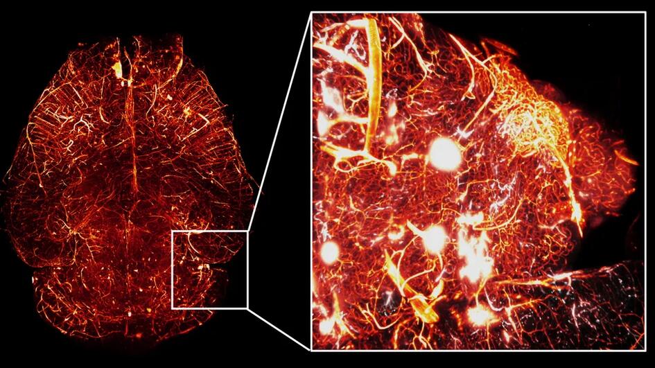
Very severe cases of malaria can cause lasting brain damage or prove fatal. Researchers from the Bernhard Nocht Institute for Tropical Medicine (BNITM), the University Medical Center Hamburg-Eppendorf (UKE) and the German Center for Infection Research (DZIF) investigated in collaboration with international partners how brain damage occurs in malaria at the cellular level. In mice, they found that the protein RIPK1, a regulator of inflammatory and cell death processes, contributes to damage to the cells lining the brain’s blood vessels. Findings from human brain tissue and genetic data from malaria patients also pointed to this protein. The results have been published in the journal Science Advances.
Malaria remains one of the deadliest infectious diseases worldwide. According to World Health Organization (WHO) estimates, there were over 280 million cases in 2024, with 610,000 people dying from the parasitic infection. 95 per cent of deaths occur in sub-Saharan Africa, where children under the age of five are particularly affected. Prompt treatment is crucial. Nevertheless, some patients die from malaria even when treated with medication. One of the most serious complications is cerebral malaria, which is characterized by a deep coma. Around 25 to 30 per cent of children who survive cerebral malaria are left with long-term impairments, for example in motor skills, learning or memory.
“We wanted to understand what happens at the cellular level in cerebral malaria. This understanding is essential for developing future drug therapies aimed at protecting the brain. We wanted to identify target structures in the brain that active substances can act upon to prevent or minimize the neurological effects of the infection,” says PD (Privatdozent, a habilitated lecturer) Dr. Thomas Jacobs, head of the Protozoan Immunology research group at BNITM and scientist in the research area Malaria and Neglected Tropical Diseases of the DZIF. Jacobs led the study together with Prof. Dr. Manuel A. Friese from the Institute of Neuroimmunology and Multiple Sclerosis at the UKE.
Focus on blood vessels
In cerebral malaria, red blood cells infected with the parasite Plasmodium falciparum can adhere on the walls of the smallest blood vessels in the brain, disrupting blood flow. Inflammatory reactions also occur and blood clotting is impaired. The brain may swell and small hemorrhages can develop. The focus here is on the endothelial cells, which line the inside of the blood vessels and form an essential part of the blood-brain barrier. This barrier regulates the exchange of substances between the blood and the brain.
The researchers wanted to find out which cellular mechanisms damage the endothelial cells in cerebral malaria. To this end, they combined an established mouse model and cell culture experiments with human tissue samples, brain imaging and an analysis of existing genetic data. The mouse model replicates important aspects of vascular damage, but differs from human malaria in terms of key disease mechanisms.
The RIPK1 protein as a potential target
To investigate the processes in the endothelial cells, the team infected mice with the malaria parasite Plasmodium berghei. The researchers then analysed messenger RNAs bound to the protein factories of the endothelial cells. These molecules contain the blueprints for proteins. The analysis revealed that gene programmes linked to cell death processes were increasingly active. The researchers also investigated CD8-positive T cells, a group of immune cells. In the mouse model, these cells contribute to vascular damage. When these cells were removed, the cell death programmes in the endothelial cells were attenuated. By analysing the RNA data from both cell types together, the team could infer possible signalling links between them. This brought RIPK1, a protein involved in several signalling pathways that regulate inflammation and cell death, into focus.
“The RIPK1 protein is fascinating because it plays a part in determining whether a cell lives or dies. Under inflammatory conditions, its enzymatic activity can contribute to cell death. That is why we wanted to test whether inhibiting the activity of RIPK1 could protect vascular cells,” explains first author Dr. Alexandros Hadjilaou, a researcher at BNITM and DZIF and a physician at the UKE.
In brain sections from infected mice, the researchers found increased levels of markers for various cell death processes. In human tissue, they investigated a different question: in eight children from Malawi who had died of cerebral malaria, the endothelial cells showed stronger signals of activated RIPK1 than in eight individuals who had died of other severe brain diseases and did not have malaria. This finding supports a possible role for the RIPK1 signalling pathway in humans. However, this tissue analysis does not prove that CD8-positive T cells are causally linked to RIPK1 activation.
The researchers also analysed existing genetic data. They included various forms of severe malaria in their analyses, not just the cerebral form. This revealed associations in humans between variants in genes involved in programmed cell death, including those in the RIPK1 signalling network, and severe malaria. These genetic associations provide further evidence that RIPK1 plays an important role. However, they do not prove that the disease mechanism operates via RIPK1 signalling pathways.
Longer survival in a mouse model
To test RIPK1 as a potential target, the researchers conducted experiments in genetically modified mice. The genetic modification silenced RIPK1 enzyme activity specifically in the endothelial cells of the brain. Following malaria infection, these mice survived longer than the control animals, but the modification did not prevent a fatal outcome. In a separate experimental group, the team inhibited RIPK1 using GNE684, an experimental compound that can also act outside the cerebral blood vessels. These mice also survived longer than their control counterparts. Treatment began as early as the first day after infection, i.e. before the onset of neurological disease. Both interventions prolonged survival. The researchers also measured the parasite load in the blood of the two mouse experimental groups; it remained comparable to that in the control animals.
Prospects for future drug development
“Our experiments show that RIPK1 contributes to damage to the brain’s blood vessels in the mouse model. This means we have identified a potential starting point for the further development of complementary treatments,” says Jacobs.
The aim is combined treatments that, alongside medicines against the parasite infection, include active substances that protect the brain. It remains an open question whether RIPK1 inhibition in patients improves survival following a malaria infection or prevents subsequent neurological damage. Further studies are needed to answer this question. Such a treatment would be intended as a complement to drugs targeting the malaria parasites. The next step is to investigate whether administering it at a later stage, together with standard malaria treatment, is effective and well-tolerated.
Researchers from eight countries across four continents took part in the study. The work was supported by the German Center for Infection Research (DZIF) and funded, amongst others, by the former Federal Ministry of Education and Research (BMBF) via DZIF, the Else Kröner-Fresenius Foundation, the German Research Foundation (DFG) via the Collaborative Research Center (CRC) 1648, the Werner Otto Foundation, and through institutional funding from the UKE.
An explainer video for the study can be found here: https://ripk1.hadjilaou.com/
Source: Press release of the Bernhard Nocht Institute for Tropical Medicine


